小分子融合肽TAT-KIR调节创伤性脑损伤后星形胶质细胞激活加速神经功能修复

张厚莹 程立雪 冯韵然 王心怡 吴晗溪 曹为众 杨舒涵 孙浩然 舒章 马凯歌 胡晓宣

解剖学报 ›› 2026, Vol. 57 ›› Issue (4) : 391-399.

PDF(1382 KB)
欢迎访问《解剖学报》官方网站!今天是 English
PDF(1382 KB)
解剖学报 ›› 2026, Vol. 57 ›› Issue (4) : 391-399. DOI: 10.16098/j.issn.0529-1356.2026.04.001
神经生物学

小分子融合肽TAT-KIR调节创伤性脑损伤后星形胶质细胞激活加速神经功能修复

  • 张厚莹程立雪冯韵然1,2 王心怡吴晗溪曹为众杨舒涵孙浩然舒章马凯歌1* 胡晓宣1,3 *

作者信息 +

Small-molecule fusion peptide TAT-KIR improving neurofunctional recovery by regulating astrocytes activation in mice after traumatic brain injury

  • ZHANG Hou-ying1, CHENG Li-xue1, FENG Yun-ran1,2, WANG Xin-yi1, WU Han-xi1, CAO Wei-zhong1, YANG Shu-han1, SUN Hao-ran1, SHU Zhang1, MA Kai-ge1*, HU Xiao-xuan1,3 *
Author information +
文章历史 +

摘要

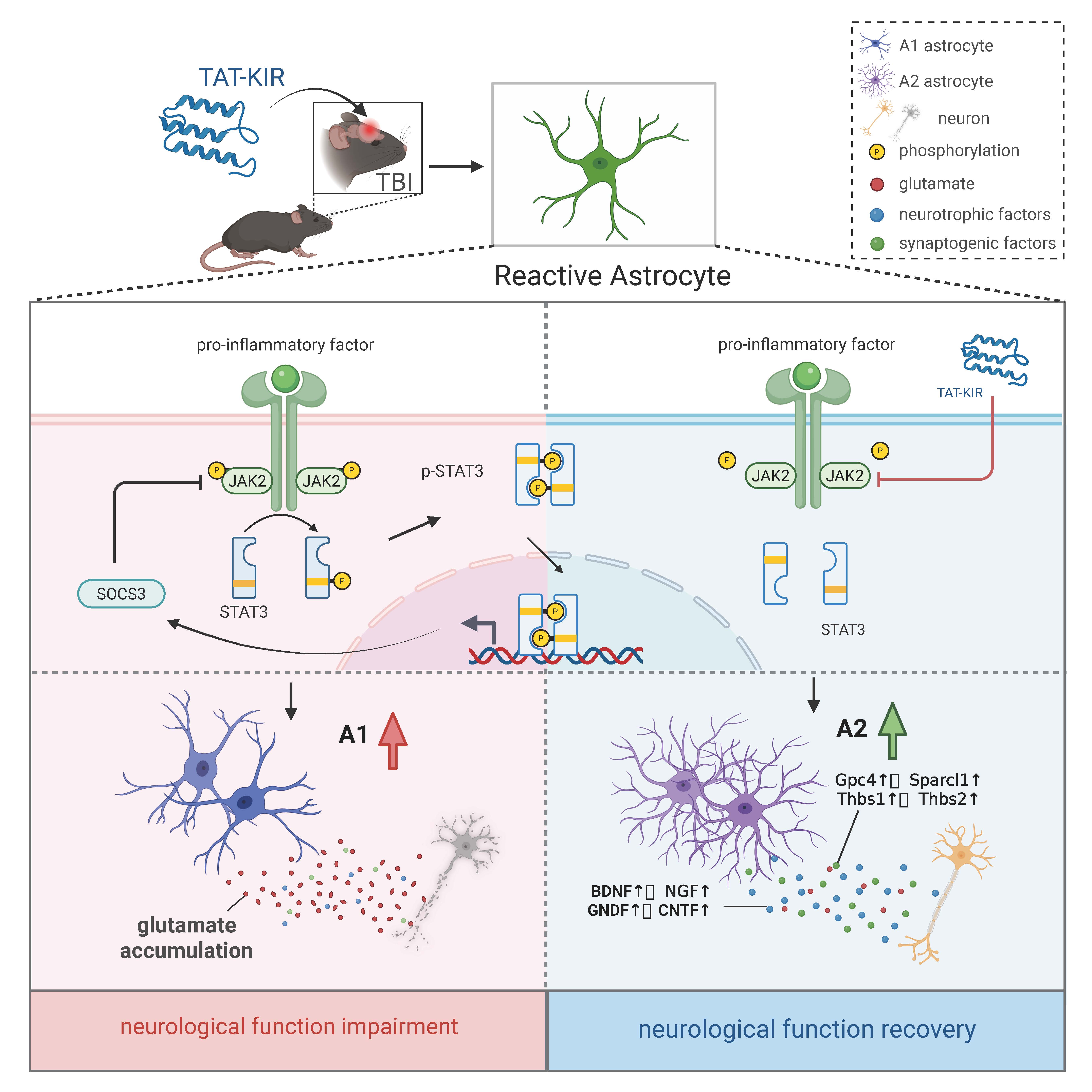
目的 探讨创伤性脑损伤(TBI)后小分子融合肽转录反式激活因子(TAT)-细胞因子信号抑制物3(SOCS3)蛋白的激酶抑制区(KIR)调节星形胶质细胞激活,促进损伤修复的作用。方法 将2月龄雄性C57BL/6小鼠随机分为假手术组(sham)、损伤组(TBI)和治疗组(TAT-KIR),每组15只。TBI组使用Feeney自由落体法构建TBI模型,TAT-KIR组在TBI后立即原位注射TAT-KIR,sham组只开颅不打击。TBI后1 d Western blotting检测小鼠皮质损伤区及周围组织JAK2/STAT3通路激活情况;TBI后1、3、7及14 d,NSS和Beam walking评估小鼠运动功能,免疫荧光染色检测星形胶质细胞和神经元数量,并使用Sholl analysis对星形胶质细胞形态进行分析;TBI后3 d和7 d,Real-time PCR检测星形胶质细胞A1型标志物C3、H2-D1、Serping1和H2-T23和A2型标志物转谷氨酰胺酶1(Tgm-1)、心肌营养素样细胞因子 1(Clcf1)、S100 钙结合蛋白 A10 (S100a10) 和CD109基因表达及相关因子水平,ELISA检测局部脑组织谷氨酸含量。结果 TAT-KIR治疗组小鼠激活的JAK2/STAT3信号通路被抑制,皮质损伤区反应性星形胶质细胞增生减弱且分支变少;C3、H2-D1、Serping1、H2-T23(A1型)表达下调,Tgm-1、Clcf1、CD109、S100a10(A2型)表达上调;促突触生成因子(Gpc4、Sparcl1、Thbs1、Thbs2)和神经营养因子[脑源性神经营养因子(BDNF)、睫状神经营养因子(CNTF)、胶质细胞源性神经营养因子(GDNF)、神经营养素-3 (NT3)、神经生长因子(NGF)] mRNA表达水平升高,谷氨酸转运蛋白1(GLT-1)和谷氨酸/天冬氨酸转运蛋白(GLAST)表达增多,皮质损伤区组织中谷氨酸含量降低;随恢复时间增加,TBI小鼠皮质损伤区神经元丢失明显减轻,NSS评分降低,平衡木移动距离增加。结论 TBI急性期给予TAT-KIR,可有效减弱星形胶质细胞反应性增生,增加A2型细胞比例和神经保护功能,加速小鼠神经功能恢复。

Abstract

ObjectiveTo investigate the therapeutic potential of the small-molecule fusion peptide trans-activator of transcription(TAT)-kinase inhibitory region(KIR)of cytokine signaling 3(SOCS3) protein in mice by modulating astrocyte activation after traumatic brain injury (TBI). Methods Male C57BL/6 mice (2-month-old) were randomly divided into three groups, sham group (sham), TBI group (TBI), and TAT-KIR group (TAT-KIR), and each group had 15 mice. The TBI group received traumatic brain injury using Feeney’s free-fall method, the TAT-KIR group was injected with TAT-KIR in situ right after TBI, the sham group underwent craniotomy without impact. Western blotting was used to assess the activation of JAK2/STAT3 pathway at day 1 post-TBI. Immunofluorescence staining was employed to evaluate the astrocyte activation and the neuron loss at day 1, 3, 7, and 14 post-TBI, and Sholl analysis was used to analyze the morphology of astrocytes. Meanwhile, neurological severity score (NSS) and beam walking tests were used to evaluate motor function. At day 3 and 7 post-TBI, Real-time PCR was performed to measure the mRNA expression levels of C3, H2-D1, Serping1, H2-T23 (A1-astrocyte) and transglutaminase 1(Tgm-1), cardiotrophin-like cytokine factor 1(Clcf1), S100 calcium-binding protein A10(S100a10), CD109 (A2-astrocyte), and the cytokines secreted by activated astrocytes. ELISA was used to detecte the amount of glutamate in injured cortex. Results In the TAT-KIR treated group, TBI-induced activation of the JAK2/STAT3 signaling pathway and astrogliosis were suppressed. The branches of activated astrocytes decreased. The mRNA level of C3, H2-D1, Serping1, H2-T23 were downregulated, while the mRNA level of Tgm-1, Clcf1, CD109, S100a10 were upregulated. At the same time, the synaptogenic factors, including Gpc4, Sparcl1, Thbs1, Thbs2, and the neurotrophic factors, such as brain-derived neurotrophic factor (BDNF), ciliary neurotrophic factor (CNTF), glial cell line-derived neurotrophic factor (GDNF), neurotrophin-3 (NT3), nerve growth gactor (NGF) were significantly upregulated. The expression of glutamate transporters, including glutamate transporter 1(GLT-1)and glutamate/aspartate transporter(GLAST)increased, while the amount of extracellular glutamate decreased. The neuron loss of TBI mice gradually reduced with the time. NSS scores decreased and the distance of beam walking significantly increased. Conclusion Administration of TAT-KIR during the acute phase of TBI reduced astrocyte activation, upregulated the proportion of A2 astrocyte, and subsequently accelerated neurological function recovery in TBI mice.

关键词

/ "> 创伤性脑损伤;转录反式激活因子-细胞因子信号抑制物3蛋白的激酶抑制区;星形胶质细胞;神经功能修复;Feeney自由落体法;免疫荧光;小鼠

Key words

Traumatic brain injury
/ Trans-activator of transcription-kinase inhibitory region of suppressor of cytokine signaling 3 protein / Astrocyte / Neurological function restoration / Feeney’s free fall / Immunofluorescence / Mouse

引用本文

导出引用
张厚莹 程立雪 冯韵然 王心怡 吴晗溪 曹为众 杨舒涵 孙浩然 舒章 马凯歌 胡晓宣. 小分子融合肽TAT-KIR调节创伤性脑损伤后星形胶质细胞激活加速神经功能修复[J]. 解剖学报. 2026, 57(4): 391-399 https://doi.org/10.16098/j.issn.0529-1356.2026.04.001
ZHANG Hou-ying, CHENG Li-xue, FENG Yun-ran, WANG Xin-yi, WU Han-xi, CAO Wei-zhong, YANG Shu-han, SUN Hao-ran, SHU Zhang, MA Kai-ge, HU Xiao-xuan. Small-molecule fusion peptide TAT-KIR improving neurofunctional recovery by regulating astrocytes activation in mice after traumatic brain injury[J]. Acta Anatomica Sinica. 2026, 57(4): 391-399 https://doi.org/10.16098/j.issn.0529-1356.2026.04.001
中图分类号: R322.81   

参考文献

[1] Roberson SW, Patel MB, Dabrowski W, et al. Challenges of delirium management in patients with traumatic brain injury: from pathophysiology to clinical practice [J]. Curr Neuropharmacol, 2021, 19(9): 1519-1544.
[2] Karthigeyan M, Gupta SK, Salunke P, et al. Head injury care in a low- and middle-income country tertiary trauma center: epidemiology, systemic lacunae, and possible leads [J]. Acta Neurochir (Wien), 2021, 163(10): 2919-2930.
[3] Du M, Wu C, Yu R, et al. A novel circular RNA, circIgfbp2, links neural plasticity and anxiety through targeting mitochondrial dysfunction and oxidative stress-induced synapse dysfunction after traumatic brain injury [J]. Mol Psychiatry, 2022, 27(11): 4575-4589.
[4] Janowitz T, Menon DK. Exploring new routes for neuroprotective drug development in traumatic brain injury [J]. Sci Transl Med, 2010, 2(27): 27rv1.
[5] Simon DW, McGeachy MJ, Bay?r H, et al. The far-reaching scope of neuroinflammation after traumatic brain injury [J]. Nat Rev Neurol, 2017, 13(3): 171-191.
[6] Yuan M, Wu H. Astrocytes in the traumatic brain injury: the good and the bad [J]. Exp Neurol, 2022, 348: 113943.
[7] Hinkle JT, Dawson VL, Dawson TM. The A1 astrocyte paradigm: new avenues for pharmacological intervention in neurodegeneration [J]. Mov Disord, 2019, 34(7): 959-967.
[8] Villarreal A, Vidos C, Monteverde Busso M, et al. Pathological neuroinflammatory conversion of reactive astrocytes is induced by microglia and involves chromatin remodeling [J]. Front Pharmacol, 2021, 12: 689346.
[9] Cieri MB, Villarreal A, Gomez-Cuautle DD, et al. Progression of reactive gliosis and astroglial phenotypic changes following stab wound-induced traumatic brain injury in mice [J]. J Neurochem, 2023, 167(2): 183-203.
[10] Miyamoto N, Magami S, Inaba T, et al. The effects of A1/A2 astrocytes on oligodendrocyte linage cells against white matter injury under prolonged cerebral hypoperfusion [J]. Glia, 2020, 68(9): 1910-1924.
[11] Cieri MB, Ramos AJ. Astrocytes, reactive astrogliosis, and glial scar formation in traumatic brain injury [J]. Neural Regen Res, 2025, 20(4): 973-989.
[12] Yu G, Zhang Y, Ning B. Reactive astrocytes in central nervous system injury: subgroup and potential therapy [J]. Front Cell Neurosci, 2021, 15: 792764.
[13] Oliva AA Jr, Kang Y, Sanchez-Molano J, et al. STAT3 signaling after traumatic brain injury [J]. J Neurochem, 2012, 120(5): 710-720.
[14] Zhao JB, Zhang Y, Li GZ, et al. Activation of JAK2/STAT pathway in cerebral cortex after experimental traumatic brain injury of rats [J]. Neurosci Lett, 2011, 498(2): 147-152.
[15] Maas AIR, Menon DK, Manley GT, et al. Traumatic brain injury: progress and challenges in prevention, clinical care, and research [J]. Lancet Neurol, 2022, 21(11): 1004-1060.
[16] Mureanu IA, Grad DA, Mureanu DF, et al. Evaluation of post-traumatic stress disorder (PTSD) and related comorbidities in clinical studies [J]. J Med Life, 2022, 15(4): 436-442.
[17] Hsueh SC, Parekh P, Batsaikhan B, et al. Targeting neuroinflammation: 3-monothiopomalidomide a new drug candidate to mitigate traumatic brain injury and neurodegeneration [J]. J Biomed Sci, 2025, 32(1): 57.
[18] Alhadidi QM, Bahader GA, Arvola O, et al. Astrocytes in functional recovery following central nervous system injuries [J]. J Physiol, 2024, 602(13): 3069-3096.
[19] Wang X, Li X, Zuo X, et al. Photobiomodulation inhibits the activation of neurotoxic microglia and astrocytes by inhibiting Lcn2/JAK2-STAT3 crosstalk after spinal cord injury in male rats [J]. J Neuroinflammation, 2021, 18(1): 256.
[20] Liddelow SA, Guttenplan KA, Clarke LE, et al. Neurotoxic reactive astrocytes are induced by activated microglia [J]. Nature, 2017, 541(7638): 481-487.
[21] Chen Y, Qin C, Huang J, et al. The role of astrocytes in oxidative stress of central nervous system: a mixed blessing [J]. Cell Prolif, 2020, 53(3): e12781.
[22] Zhang H, Zhang X, Chai Y, et al. Astrocyte-mediated inflammatory responses in traumatic brain injury: mechanisms and potential interventions [J]. Front Immunol, 2025, 16: 1584577.
[23] Mutoji KN, Sun M, Nash A, et al. Anti-inflammatory protein TNFα-stimulated gene-6 (TSG-6) reduces inflammatory response after brain injury in mice [J]. BMC Immunol, 2021, 22(1): 52.
[24] Min H, Choi B, Jang YH, et al. Heme molecule functions as an endogenous agonist of astrocyte TLR2 to contribute to secondary brain damage after intracerebral hemorrhage [J]. Mol Brain, 2017, 10(1): 27.
[25] Boghdadi AG, Teo L, Bourne JA. The neuroprotective role of reactive astrocytes after central nervous system injury [J]. J Neurotrauma, 2020, 37(5): 681-691.

基金

国家自然科学基金(82171389);中央高校基本科研业务费专项资金(xzy012025169)

PDF(1382 KB)

Accesses

Citation

Detail

段落导航
相关文章

/